
Infection with extensive gas formation due to myonecrosis, also 3,4,6,8 The muscle is typically edematous, partially necrotic, and can contain multiple abscesses. Theīuttock and thigh are the most commonly affected locations, and Staphylococcus aureus ( S. Immunosuppression are also at increased risk for muscle infection. 3,4,6ĭiabetic patients and other patients with vascular compromise or Recognized to be at high risk for deep muscular infection. The tropics more recently, patients infected with HIV have been Process was most commonly encountered in children and in patients from Of muscle is referred to as “pyomyositis.” In the past, this disease
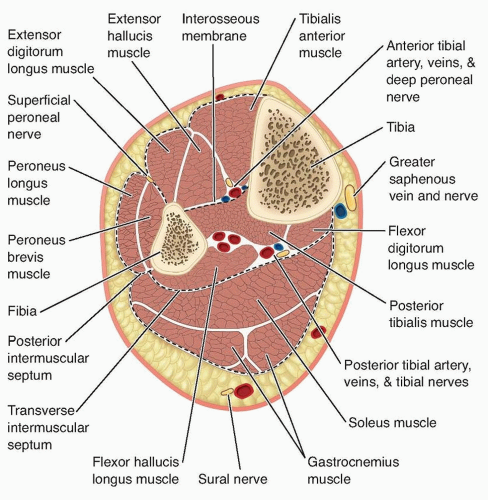
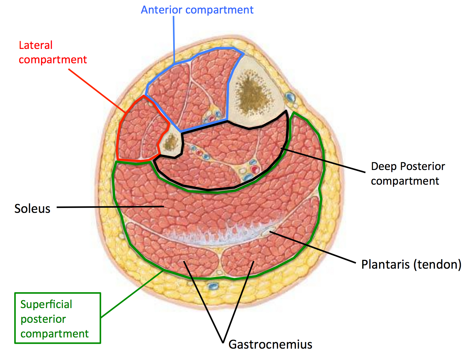
#Compartments of leg radiology skin
Tissue and skin inflammation are generally present. The thick, irregular wall enhancesĪfter administration of intravenous gadolinium (Figure 2). The proteinaceous granulation tissue on the inner margin may show Images and high signal intensity on T2-weighted (T2W) images, though The typicalĪbscess shows central areas of low signal intensity on T1-weighted (T1W) 4Ībscesses have variable signal characteristics on MRI. Both radiographs and CTĬan demonstrate retained foreign bodies that may be the cause ofĪbscess, although CT is more sensitive. CT is particularly useful forĭetecting gas present within the abscess cavity. Inflammatory changes in the soft tissues adjacent to the abscess may

Margins that enhance after the administration of intravenous contrast. 6 The CT appearance ofĪn abscess is a heterogenous fluid collection with thick irregular Radiographs, an abscess appears as a nonspecific soft-tissue mass Ībscesses are difficult to diagnose with specificity, unless gas bubbles Soft-tissue infection can result in the formation of an abscess. Should be undertaken if the imaging findings are equivocal. Definitiveĭiagnosis of necrotizing fasciitis is made by fascial biopsy, which Perifascial gas and areas of tissue necrosis (Figure 1). Resonance imaging (MRI) are useful for noninvasive identification of 5,6 Computed tomography (CT) and magnetic Planes, affording specific radiographic diagnosis, but this finding has Occasional cases demonstrate small gas bubbles adjacent to fascial 5 Necrotizing fasciitis is a surgical emergency, so rapid and accurate diagnosis is imperative. Necrotizing fasciitis is a fulminant form of septic fasciitisĪssociated with rapid spread of infection and prominent tissue necrosis. Refers to infection that has extended to involve the fibrous fascia Superficial to the superficial fascia, the term “septic fasciitis” Occasionally, small fluidĬollections may be present in the subdermal areas or superficial to the 4 OnĬross-sectional imaging, cellulitis results in thickening of the skinĪnd the septae in the subcutaneous tissue. Soft-tissue swelling, and obliteration of fat planes. Radiographic findings of cellulitis include skin thickening, nonspecific Immunosuppression, recent trauma, and retained foreign bodies. Insufficiency, soft-tissue ulcer (often secondary to diabetes), Superficial compartments and working progressively deeper to the boneĬellulitis, fasciitis, soft -tissue abscess, and pyomyositisĬellulitis is an acute infectious process limited to the skin and subcutaneous tissues. The various layers of musculoskeletal tissue, starting in the ThisĪrticle will review the imaging characteristics of infection involving Osteomyelitis representing the most severe and deepest level. Of such tissue layers may coexist or develop sequentially, with 1,2 Various anatomic planes can be involved (Table 1). Mortality, making rapid and accurate diagnosis crucial. Delayed diagnosis can lead to significant morbidity and Infection, the organism involved, and the underlying health of the


Treatment and prognosis depend upon the site of Infection of the musculoskeletal system is commonly encountered inĬlinical practice. Is a Professor of Radiology and the Residency Program Director, in theĭepartment of Radiology, University of California, San Diego, San Diego,Ĭertain images and text from this publication were presented as an educational exhibit at the RSNA 2009 meeting. Pathria is a Professor of Radiology, and Dr.


 0 kommentar(er)
